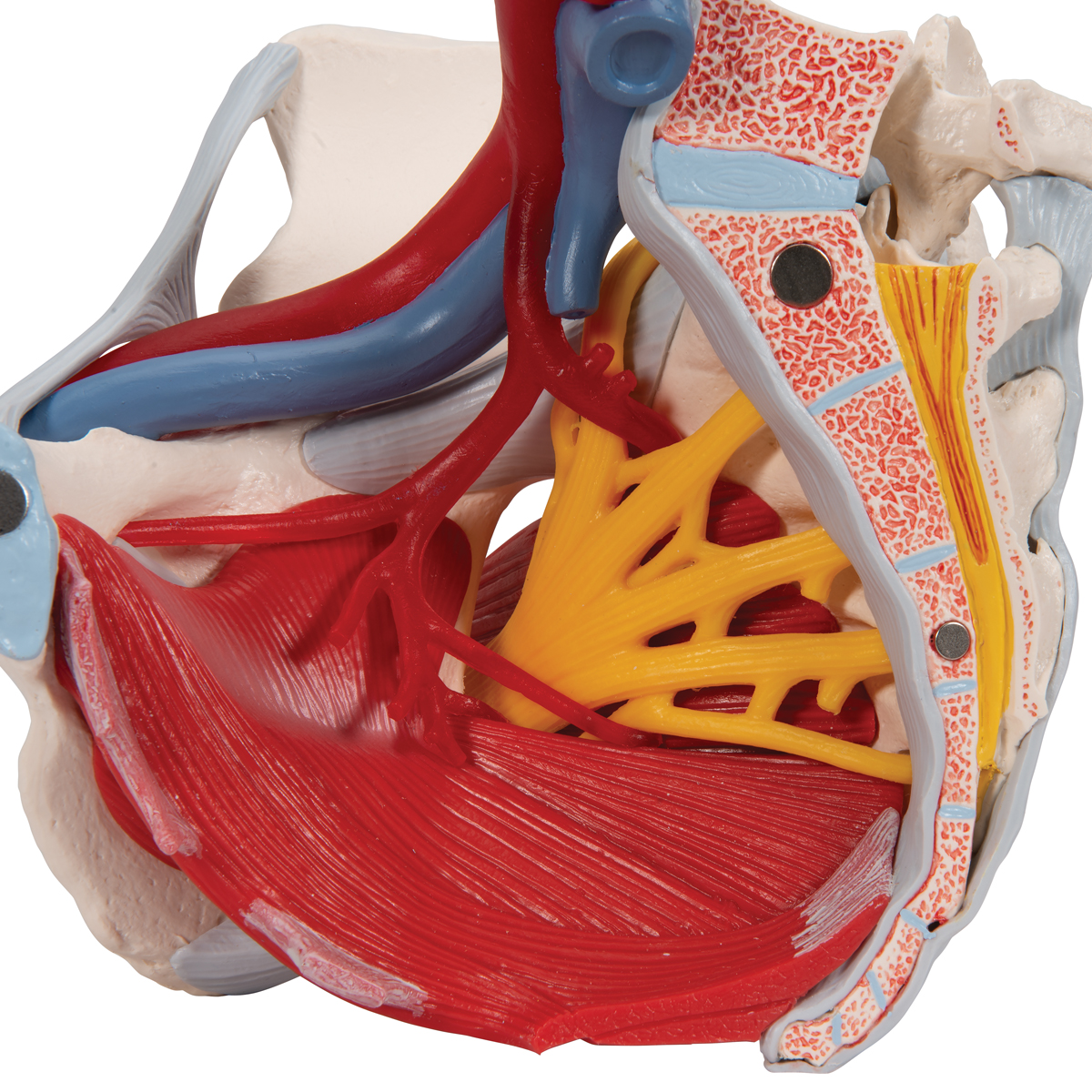
Human Anatomy Muscles Pelvis - Human Pelvis Muscle Bone Anatomy 3D Model MAX OBJ 3DS FBX ... : These muscles origin in continuity from the body of the pubis.
byAdmin-
0
Human Anatomy Muscles Pelvis - Human Pelvis Muscle Bone Anatomy 3D Model MAX OBJ 3DS FBX ... : These muscles origin in continuity from the body of the pubis.. These muscles, including the gluteus maximus and the hamstrings, extend the thigh at the hip in support of the body's weight and propulsion. For didactic purposes and practice, we labeled one tenth of the possible structures to not. The floor of the pelvis is formed by the two muscles named levator ani and coccygeus. Pelvis bones and the ligaments front on and rear view. A variably thick muscular membrane called a diaphragm coccygeus and levator ani muscles (iliococcygeus, puborectalis the lower part of the pelvis is sealed off by a muscular diaphragm and perineal membrane known as the pelvic floor.
The floor of the pelvis is formed by the two muscles named levator ani and coccygeus. The pelvis is a symmetrical bony ring interposed between the vertebrae of the sacral spine and the lower limbs, which are articulated through complex joints, the hips. Muscle movements, types, and names. Of human anatomy and different types of motion, inspiring more realistic and energetic figurative art. Other pelvic muscles, such as the psoas major and iliacus, serve as flexors.
Image result for ann frye paper pelvic floor | Female ... from i.pinimg.com Anatomy atlases, the anatomy atlases logo, and a digital library of anatomy information are all trademarks of michael p. You can click the links in the image, or the links below the image to find out more information on any muscle group. Pelvic diaphragm the muscles with the covering fascia. Human the muscular system consists of the skeletal muscles and their associated structures. Anatomy of the muscular system. Anatomy of the human body henry gray contents i. Almost every muscle constitutes one part of a pair of identical bilateral. Is a registered osteopath, lecturer and author and is discussing the anatomy and function of the human pelvis and includes the si joint and lumbar spine.
Anatomy of a human body we study anatomy.
Functional anatomy of the pelvis, sacroiliac joint and lumbar spine. The information contained in anatomy atlases is not a substitute for the medical care and advice of your physician. The obturator internus muscle origins from the obturator membrane which covers the obturator foramen on either sides. Blood supply functions • to support the pelvic organs • to maintain the intra abdominal pressure by reflexly responding tomits changes. Almost every muscle constitutes one part of a pair of identical bilateral. This is a table of skeletal muscles of the human anatomy. Attached to the pelvis are muscles of the buttocks, the lower back, and the thighs. Find the best weight lifting exercises that target each muscle or groups of muscles. Urogenital diaphragm main structures ischial tuberosity pubic symphysis coccyx sacrotuberous ligament ischipubic ramus. Adj основной cartilage ˈkɑːrtɪlɪdʒ n хрящ pelvis 'pelvis n таз ligament ˈlɪɡəmənt n связка substance. The visible human project is a fantastic tool that allows you to view almost all anatomical structures of the body. Pelvis bones and the ligaments front on and rear view. Gutman, md objectives understand pelvic anatomy organs and structures of the female pelvis vascular supply neurologic supply epicranius anatomy and physiology 121:
Learn about anatomy muscles pelvis with free interactive flashcards. The muscles of the pelvis, hip and buttock anatomical chart shows how each muscle in this area of the body works with the others, and the various minor systems within the major ones. You can click the links in the image, or the links below the image to find out more information on any muscle group. Find the best weight lifting exercises that target each muscle or groups of muscles. Functional anatomy of the pelvis, sacroiliac joint and lumbar spine.
Anatomical Teaching Models | Plastic Human Pelvic Models ... from www.3bscientific.com The pelvis is a symmetrical bony ring interposed between the vertebrae of the sacral spine and the lower limbs, which are articulated through complex joints, the hips. Human the muscular system consists of the skeletal muscles and their associated structures. Muscle movements, types, and names. It supports the spinal column and connects the upper body to the lower extremities. Microscopic anatomy of skeletal muscle. Innervation of the female levator ani muscles. Find the best weight lifting exercises that target each muscle or groups of muscles. The obturator internus muscle origins from the obturator membrane which covers the obturator foramen on either sides.
Human anatomy » musculoskeletal system » the muscles of the abdomen, lower back located pelvic girdle, the muscles of this region play a critical role in protecting the delicate vital organs extending across the anterior surface of the body from the superior border of the pelvis to the inferior.
It supports the spinal column and connects the upper body to the lower extremities. (1) the obturator internus and the piriformis, which are muscles of the lower extremity, and will it arches beneath the obturator vessels and nerve, completing the obturator canal, and at the front of the pelvis is attached to the back of the. Anatomy atlases, the anatomy atlases logo, and a digital library of anatomy information are all trademarks of michael p. Anatomy of a human body we study anatomy. This is a table of skeletal muscles of the human anatomy. Included within the chart are gorgeous illustrations of the pelvic diaphragm, sphincter muscles, gluteus maximus. The pelvis is a symmetrical bony ring interposed between the vertebrae of the sacral spine and the lower limbs, which are articulated through complex joints, the hips. 12 frolich, human anatomy, pelvis i the pelvic floor muscular floor and sphinchters transverse perineal m. Blood supply functions • to support the pelvic organs • to maintain the intra abdominal pressure by reflexly responding tomits changes. Architectural differences in the bony pelvis of women with and without pelvic floor disorders. Only the most important muscles are described here because it is beyond our scope to describe the hundreds of skeletal muscles of the human body. The floor of the pelvis is formed by the two muscles named levator ani and coccygeus. Innervation of the female levator ani muscles.
The muscles of the pelvis, hip and buttock anatomical chart shows how each muscle in this area of the body works with the others, and the various minor systems within the major ones. Anatomy of the muscular system. The floor of the pelvis is made up of the muscles of the pelvis, which support its contents and maintain urinary there are many organs that sit in the pelvis, including much of the urinary system, and lots of the in this section, learn more about the anatomy of the pelvis, and the structures located within it. The visible human project is a fantastic tool that allows you to view almost all anatomical structures of the body. Cross the hip joint onto the thigh/leg 3.
Anatomical Teaching Models | Plastic Human Pelvic Models ... from www.3bscientific.com Muscle movements, types, and names. You can click the links in the image, or the links below the image to find out more information on any muscle group. Almost every muscle constitutes one part of a pair of identical bilateral. Key facts about the muscles of the pelvic floor. Human anatomy » musculoskeletal system » the muscles of the abdomen, lower back located pelvic girdle, the muscles of this region play a critical role in protecting the delicate vital organs extending across the anterior surface of the body from the superior border of the pelvis to the inferior. Urogenital diaphragm main structures ischial tuberosity pubic symphysis coccyx sacrotuberous ligament ischipubic ramus. The muscle begins near the asis of the pelvis directly above the acetabulum (hip socket). Learn about anatomy muscles pelvis with free interactive flashcards.
There are around 650 skeletal muscles within the typical human body.
Gutman, md objectives understand pelvic anatomy organs and structures of the female pelvis vascular supply neurologic supply epicranius anatomy and physiology 121: This is a table of skeletal muscles of the human anatomy. The muscles within the pelvis may be divided into two groups: Innervation of the female levator ani muscles. Read and learn the following words: Pelvic diaphragm the muscles with the covering fascia. Key facts about the muscles of the pelvic floor. Discover the muscle anatomy of every muscle group in the human body. The floor of the pelvis is formed by the two muscles named levator ani and coccygeus. Included within the chart are gorgeous illustrations of the pelvic diaphragm, sphincter muscles, gluteus maximus. These muscles, including the gluteus maximus and the hamstrings, extend the thigh at the hip in support of the body's weight and propulsion. There are 26 bones in the human foot which are grouped into 7 tarsals, 5 metatarsals and 14 phalanges, for a total of 33 joints, of which 20 are actively articulated. The information contained in anatomy atlases is not a substitute for the medical care and advice of your physician.
Anatomy of a human body we study anatomy anatomy muscles pelvis. Pelvic diaphragm the muscles with the covering fascia.